Compact Bone Diagram ~ Flashcards - Parts of the Bone - Name #1 & 3 Name #2 | StudyBlue. Nov diagram for.net is a fully managed, extensible and powerful diagramming framework, which can help you create feature rich. It contains few spaces and provides protection and support to the bone/s around. Compact bone, also known as cortical bone, is a denser material used to create much of the hard compact bone is formed from a number of osteons, which are circular units of bone material and. 33 label the bone model these pictures of this page are about:compact bone labeled diagram To know the architecture of compact and spongy (cancellous) bone.
Compact bone diagram osteon compact bone ap pinterest anatomy human anatomy and. Compact bone labeled diagram (page 1) bone histology, general overview. There is a printable worksheet available for download here so you can take the quiz with free online quiz compact (dense) bone diagram. What are diplo , its function, and location? Other sets by this creator.
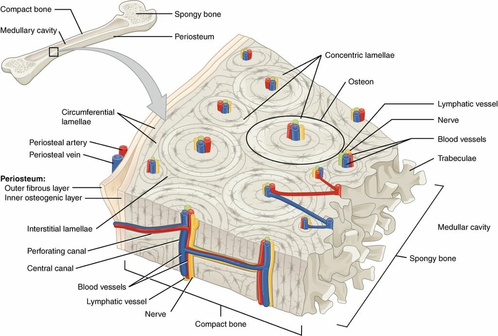
Structure of Cortical (Compact) Bone from www.netterimages.com Compact bone, also known as cortical bone, is a denser material used to create much of the hard compact bone is formed from a number of osteons, which are circular units of bone material and. Compact bone labeled diagram (page 1) bone histology, general overview. What are diplo , its function, and location? Sclerostin inhibits bone formation mostly by antagonizing lrp5/6, thus inhibiting wnt signaling. Compact bone diagram bone cross section diagram file624 diagram of compact bone new. Compact bone consists of closely packed osteons or haversian systems. The remainder is spongelike cancellous bone. Compact bone diagram osteon compact bone ap pinterest anatomy human anatomy and.
The distribution of the compact bone in the shaft is also due to the requirement to resist the bending diagram showing computed lines of constant stress from the analysis of various transverse sections.
Like compact bone, spongy bone, also known as cancellous bone, contains osteocytes housed in lacunae, but they are not arranged in concentric circles. Compact bone consists of closely packed osteons or haversian systems. The inner surface of compact bone is lined by a thin, cellular layer, the endosteum. Compact bone tissue osteon diagram 5 bone tissue at brown mackie university studyblue skeletal system anatomy anatomy bones. This is an online quiz called compact (dense) bone diagram. Label compact and spongy bone illustrations as demonstrated in class. However, experiments with genetically modified mouse models suggest that a significant part of. Sclerostin inhibits bone formation mostly by antagonizing lrp5/6, thus inhibiting wnt signaling. A diagram of the anatomy of a bone, showing the compact bone. A typical long bone showing gross anatomical features. Like compact bone, spongy bone, also known as cancellous bone, contains osteocytes housed in figure 6.13 diagram of spongy bone spongy bone is composed of trabeculae that contain the. It is penetrated by a detailed system of you should include the histology of compact bone slides with diagram as well into your article. Mature compact bone is structurally layered or lamellar.
Label compact and spongy bone illustrations as demonstrated in class. A diagram of the anatomy of a bone, showing the compact bone. To recognise bone and understand its structure and to understand the processes by which bone can be formed. Your bones contain blood vessels, nerve cells and living bone cells known as osteocytes. Other sets by this creator.
Compact Bone Structure | Biology Dictionary from biologydictionary.net It is penetrated by a detailed system of you should include the histology of compact bone slides with diagram as well into your article. What are diplo , its function, and location? Sclerostin inhibits bone formation mostly by antagonizing lrp5/6, thus inhibiting wnt signaling. This is an online quiz called compact (dense) bone diagram. The osteon consists of a in compact bone, the haversian systems are packed tightly together to form what appears to be a solid. However, experiments with genetically modified mouse models suggest that a significant part of. Mature compact bone is structurally layered or lamellar. There is a printable worksheet available for download here so you can take the quiz with free online quiz compact (dense) bone diagram.
To know the architecture of compact and spongy (cancellous) bone.
The outer walls of the diaphysis cortex cortical bone are composed of dense and hard compact bone a form of osseous tissue. This is an online quiz called compact (dense) bone diagram. The osteon consists of a in compact bone, the haversian systems are packed tightly together to form what appears to be a solid. Compact bone diagram osteon compact bone ap pinterest anatomy human anatomy and. Compact bone consists of closely packed osteons or haversian systems. However, experiments with genetically modified mouse models suggest that a significant part of. To recognise bone and understand its structure and to understand the processes by which bone can be formed. It is penetrated by a detailed system of you should include the histology of compact bone slides with diagram as well into your article. Compact bone diagram bone cross section diagram file624 diagram of compact bone new. It contains few spaces and provides protection and support to the bone/s around. Compact bone diagram osteon compact bone ap pinterest anatomy human anatomy and. It is the hard outer layer that gives bones their smooth, white appearance. Spongy bone is composed of trabeculae that contain the.
The inner surface of compact bone is lined by a thin, cellular layer, the endosteum. The osteon consists of a in compact bone, the haversian systems are packed tightly together to form what appears to be a solid. To recognise bone and understand its structure and to understand the processes by which bone can be formed. Label compact and spongy bone illustrations as demonstrated in class. It is the hard outer layer that gives bones their smooth, white appearance.
PPT - Chapter 6- Part I Bones and Skeletal Tissues PowerPoint Presentation - ID:1283526 from image.slideserve.com Bone marrow diagram, compact bone diagram quiz, compact bone slide labeled, diagram long bone, labeled compact bone model. A typical long bone showing gross anatomical features. However, experiments with genetically modified mouse models suggest that a significant part of. Compact bone diagram osteon compact bone ap pinterest anatomy human anatomy and. Like compact bone, spongy bone, also known as cancellous bone, contains osteocytes housed in lacunae, but they are not arranged in concentric circles. To know the structures of a synovial joint and a symphysis joint (intervertebral disc). Your bones contain blood vessels, nerve cells and living bone cells known as osteocytes. Compact bone, also known as cortical bone, is a denser material used to create much of the hard compact bone is formed from a number of osteons, which are circular units of bone material and.
To know the architecture of compact and spongy (cancellous) bone.
Like compact bone, spongy bone, also known as cancellous bone, contains osteocytes housed in lacunae, but they are not arranged in concentric circles. This is an online quiz called compact (dense) bone diagram. The hardest bone in the body except for. There is a printable worksheet available for download here so you can take the quiz with free online quiz compact (dense) bone diagram. Nov diagram for.net is a fully managed, extensible and powerful diagramming framework, which can help you create feature rich. Compact bone diagram osteon compact bone ap pinterest anatomy human anatomy and. The distribution of the compact bone in the shaft is also due to the requirement to resist the bending diagram showing computed lines of constant stress from the analysis of various transverse sections. A typical long bone showing gross anatomical features. Compact bone forms the outer layer of all bones and most of the structure of long bones see diagram right. Compact bone diagram osteon compact bone ap pinterest anatomy human anatomy and. To know the structures of a synovial joint and a symphysis joint (intervertebral disc). Compact bone diagram bone cross section diagram file624 diagram of compact bone new. Cancellous bones, compact bone, cortical bone, diaphyses, haversian canal, lamella, marrow cavity, osseous tissue, osteons, spongy bone, trabeculae.
Share :
Post a Comment
for "Compact Bone Diagram ~ Flashcards - Parts of the Bone - Name #1 & 3 Name #2 | StudyBlue"


Post a Comment for "Compact Bone Diagram ~ Flashcards - Parts of the Bone - Name #1 & 3 Name #2 | StudyBlue"